By: Eric Lentz, SPECTRUM Writer
Constituting one of the kingdoms of life, the fungi are a remarkable group of organisms. The fungal kingdom includes yeasts, moulds, and pathogens that cause conditions such as athlete’s foot, but fungi are perhaps best identified as the mushrooms we eat and see scattered across lawns or growing on dead wood. Despite their unique presentations above the soil, fungus mass is primarily composed of subterranean strands of cells known as hyphae (Microbiology Society 2017). Hyphae grow and branch to form a vast network of thread like strands called mycelium (Microbiology Society 2017). The mycelium from a single fungus can stretch for metres under the soil. In fact, the mycelium of a single fungus in a Michigan forest is estimated to cover a minimum of 40 acres, weigh about 100 tons, and be at least 1,500 years old, while the mycelium of another fungus in the state of Washington covers about 1,500 acres (Casselman 2007)! The hyphae secrete digestive enzymes that break down organic matter in the environment, which the fungus then absorbs (Frankland 1997). It is not just mushroom producing fungi that have this structure – the mould we see on food are also composed of hyphal strands, which collectively appear as that white fuzz covering your strawberries!
Composed of tightly packed and specialized hyphae, mushrooms are the fruiting body of a fungus and serve to house and distribute the spores of the fungus, which are analogous to plant seeds (Microbiology Society 2017). Many mushrooms consist of a stalk attached to a cap. The underside of the cap contains gills or pores, which contain the spores. Although the gilled toadstool mushroom is probably the most commonly depicted mushroom, many others such as boletes, polypores, and puffballs exist.
While walking on UTSC grounds or in the valley behind it, I often use my phone to take pictures of the variety of mushrooms and other fungi I come across. Some of the better pictures I have taken are presented in this article, as well as photos of other interesting fungi (from the Web because mine were bad-quality). Although it is difficult to identify mushrooms due to their ambiguous morphology, I have attempted to identify mushrooms where possible, using the National Audubon Society Field Guide to North American Mushrooms. Features such as habitat, colour, type of gill, type of stalk, and spore prints are commonly used to identify mushrooms. Spore prints involve putting the cap of a mushroom on a piece of paper and observing the colour and pattern of the ‘print’ that spores from the mushroom leave behind on the paper. However, it is still extremely difficult to identify mushrooms without using more advanced techniques.
Class I: Mushrooms (‘toadstools’) and Boletes
This class contains your typical mushroom. While the name ‘mushroom’ is usually used for all fruiting fungus bodies, it specifically refers to the toadstool type mushrooms with gills, such as the mushrooms we find at the grocery store. Boletes are also toadstool-like but have pores on the underside of their cap, rather than gills (Kuo 2013). The arrangement of pores in a bolete mushroom makes it look like the surface of a sponge, as pictured below.

A mushroom identified as a Two-coloured Bolete (Boletus bicolor). Notice spongy appearance of the pores on the underside of the cap (Kuo 2015).
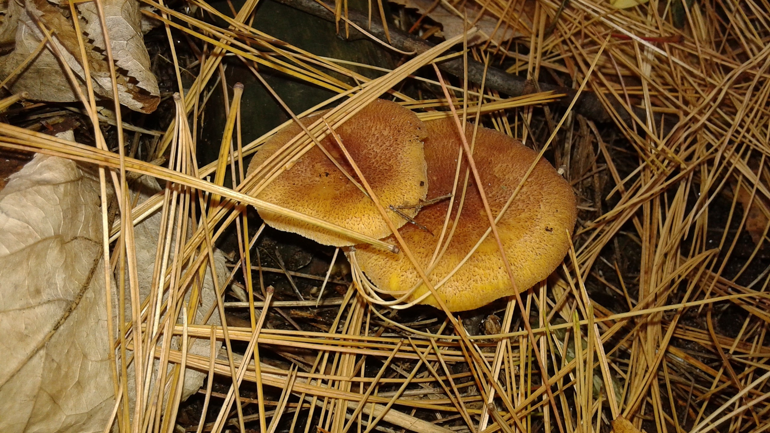
Unidentified gilled mushroom (EL 2016).


Unidentified gilled mushrooms (EL 2016).

Mushrooms identified as the Oyster Mushroom (Pleurotus ostreatus). Can also see Tinder Polypore (Fomes fomentarius) circled in red (EL 2016).
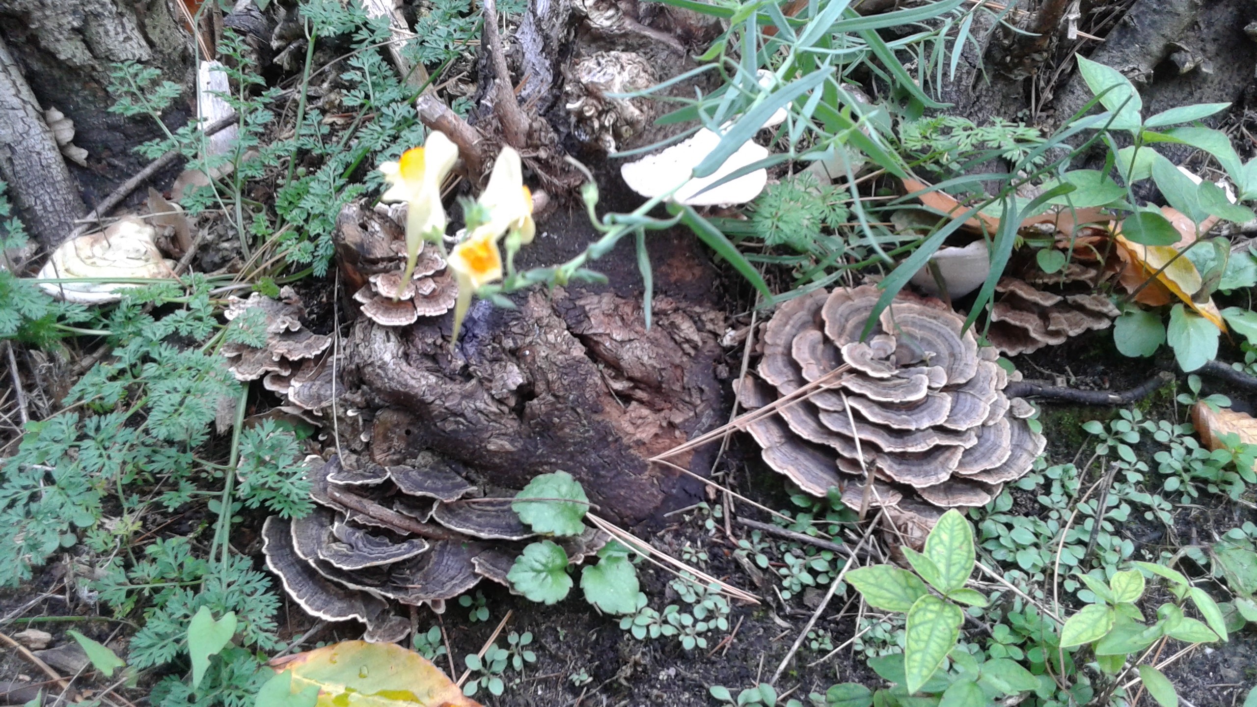
Class II: Polypores and Shelf fungi
Polypores differ morphologically from ‘toadstool’ mushrooms in that they have pores/tubes on their underside, are less soft and fleshy and are tougher and more shelf-like (Lincoff 2006). Although boletes also have pores, they are stalked and contain more flesh, unlike many polypores. Although ground polypores are mostly pictured here, they are are often seen jutting out from dead and living trees, as is the case with the Tinder Polypore below.

Possibly Turkey-tail polypores (Trametes versicolor) and/or Dye Polypore (Phaeolus schweinitzii) shown above and below (EL 2016).

Class III: Puffballs
Puffballs are spherical fungi, sometimes stalked, with spore filled interiors. The spores are exposed and are carried off by wind or rain when the outer surface of the puffball is punctured (or when someone decides to stomp on one). They can be extremely massive, with some reportedly having diameters of over 50 cm (Kuo 2008)! I was very impressed with the size of the puffballs I found in the forest behind UTSC, pictured here.
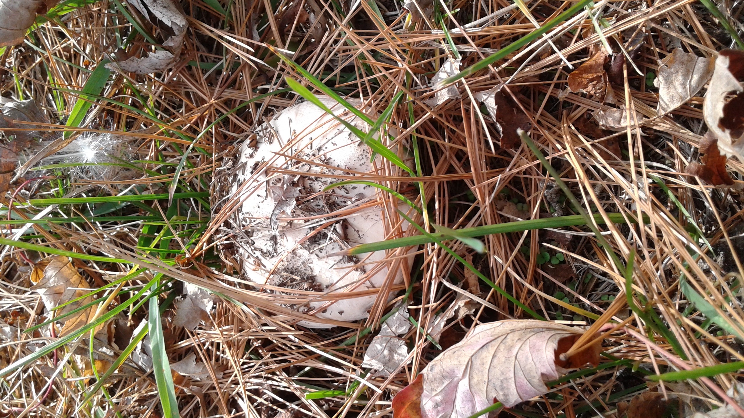
Identified to be the Western Lawn Puffbowl (Vascellum pratense) (EL 2016)

Large puffballs, speculated to be the Purple-spored puffball (Calvatia cyathiformis). My 30cm shoe is shown beside two of them as a comparison of size. Saw 10 or more of these in the middle of the forest behind UTSC. (EL 2016)

Class IV: Cup fungi, slime moulds and other fungi
I grouped these into a miscellaneous category. I have seen these fungi near UTSC but did not take good quality pictures of them, so with the exception of the Ochre Spreading Tooth fungi, I did not take these pictures but found pictures that looked similar to the fungi I saw.

Clockwise, from top right: Species identified as Ochre Spreading Tooth (Steccherinum ochraceum), Chocolate Tube Slime (Stemonitis splendens), Orange Peel fungi (Aleuria aurantia), and Bear’s Head Tooth (Hericium coralloides), Yellow-tipped coral (Ramaria formosa).
(EL 2016; Toronto Wildlife 2015; Patterson 2015; Ramer et al. n.d.; Hunter 2016)
References
Casselman, Anne (2007). “Strange but True: The Largest Organism on Earth Is a Fungus.” Scientific American. Retrieved March 2017 from: https://www.scientificamerican.com/article/strange-but-true-largest-organism-is-fungus/
Frankland, Juliet C (1997). “All you ever wanted to know about Mycelium”. NWFG Newsletter. Retrieved March 2017 from: http://fungus.org.uk/nwfg/mycapr97.htm
Hunter, Margie (2016). “Forney Ridge & Springhouse Branch Trails, August 28 & 29, 2016”. Smokies Blog. Retrieved March 2017 from: https://hikinginthesmokies.wordpress.com/2017/01/22/forney-ridge-springhouse-branch-trails-august-28-29-2016/fungi-ramaria-formosa-springhouse-branch-trail-gsmnp-august-29-2016/
Justin (2014). “Coprine: Alcohol Poisoning From Mushrooms?” Nature’s Poisons. Retrieved March 2017 from: https://naturespoisons.com/2014/04/10/coprine-alcohol-poisoning-from-mushrooms/
Kuo, M. (2015). “Boletus bicolor”. MushroomExpert.Com. Retrieved March 2017 from: http://www.mushroomexpert.com/boletus_bicolor.html
Kuo, M. (2013). “The boletes”. MushroomExpert.Com. Retrieved March 2017 from: http://www.mushroomexpert.com/boletes.html
Kuo, M. (2008). “Puffballs”. MushroomExpert.Com. Retrieved March 2017 from: http://www.mushroomexpert.com/puffballs.html
Lincoff, Gary H (2006). National Audubon Society Field Guide to North American Mushrooms. Alfred A. Knopf Inc.
Microbiology Society (2017). “Fungi.” Microbiology Online. Retrieved March 2017 from: http://microbiologyonline.org/about-microbiology/introducing-microbes/fungi
Patterson, Susan (2015). “Cup Fungi Info: What is Orange Peel Fungus”. Gardening Know How. Retrieved March 2017 from: https://www.gardeningknowhow.com/ornamental/fungus-lichen/orange-peel-fungus.htm
Ramer, Hannah; Silver, Emily; Weizmann, Limor (n.d.). “Bear’s Head Tooth”. Taste of the Wild. Retrieved March 2017 from: http://www.bio.brandeis.edu/fieldbio/Edible_Plants_Ramer_Silver_Weizmann/Pages/spp_page_Hericium_coralloides.html
Toronto Wildlife (2015). “More Chocolate Tube Slime Mould”. Toronto-wildlife.com. Retrieved March 2017 from: http://toronto-wildlife.com/Fungi/Slime_moulds/more_ct_slime.html
Hide